The Best Imaging Tests for Detecting Cancer: Their Uses and Benefits, Symptoms
Imaging tests enable doctors in taking a look at the tissues inside the body and help doctors in assessing the spread of cancerous tissue inside the body and the success of treatment. From locating the site of the tumour to assessing its severity, predicting whether a tumour is cancerous to detecting the stage of cancer and treatment plan imaging tests find wide application. There are several imaging tests used for detecting cancer and each has its benefits.
Ultrasound
Ultrasound, popularly known as sonography or ultrasonography that uses sound waves of high frequencies for creating images of the internal organs. On hitting the internal organs, the sound waves bounce back to the transducer device. The transducer converts the sound waves into pictures. The echo patterns differ for abnormal and healthy tissues assisting doctors in detecting the possibility of a tumour.
Sonography aids doctors in detecting the exact location of tumours in the body that do not show up on the x-rays. The convenience of using the ultrasound imaging test is that it is a painless procedure that can be performed quickly without prior special preparation. Doctors use sonography to differentiate between fluid-filled cysts and solid tumours based on their various echo patterns.
Magnetic Resonance Imaging (MRI)
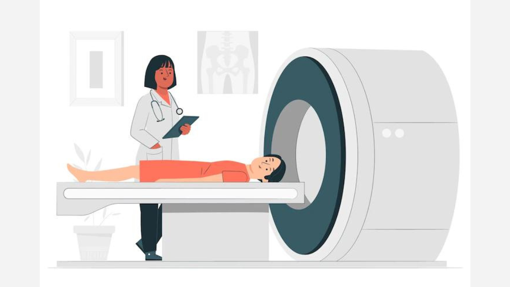
Magnetic Resonance Imaging or MRI is a popular scan to detect cancer. The imaging test utilizes magnets and radio waves for producing computer-generated detailed images of the body. Doctors use it for measuring the tumour size. The advantage of using an MRI scan is that it is safe to use on even pregnant women. Doctors use the MRI scan for identifying the nature of a tumour; whether it is non-cancerous or cancerous.
They use it for taking pictures of the different regions of the body such as the breast, abdomen, chest, spinal column and brain. MRI helps in determining the location and size of a tumour, planning the treatment and assessing the success of a treatment plan. It helps a surgeon to decide whether he should choose surgery or radiation therapy for cancer treatment.
Computed Tomography Scan (CT Scan)
Doctors use the CT scan to detect cancer inside the body in different regions such as the abdomen, neck, head, pelvis, limbs and chest. The CT scan is beneficial in knowing the exact stage of the cancerous tissue. Based on that doctors formulate the treatment plan for a patient and can predict the chances of his/her complete recovery. From CT-guided biopsies to RFA (radiofrequency ablation) CT scans find wide application.
The CT-guided biopsy is the procedure by which doctors remove tiny tissue pieces with CT scan-guided needles. Doctors choose the RFA method to treat tumours that cannot be surgically operated on. RFA can be used in conjunction with other treatment procedures such as radiation therapy, surgery, hepatic arterial infusion therapy, chemotherapy, chemoembolization and alcohol ablation. It is used in planning radiation therapy, identifying the exact location for doing the biopsy procedure and assessing the effectiveness of a treatment plan.
Mammography
The widely used scan to detect cancer of the breast is Mammography. Two kinds of mammograms; diagnostic and screening mammograms are used widely for detecting breast cancers. Doctors use the screening mammogram for detecting breast cancer at an early stage in asymptomatic women suffering from the disease.
Such mammograms are used for identifying suspicious lumps in the breast; whereas diagnostic mammograms are used for getting detailed images of the breast after doctors show some signs of cancer such as nipple discharge, breast pain or changes in shape or signs of the breast. In addition to detecting tumours, mammograms are used for detecting ductal carcinoma in situ (DCIS). Some, and not all DCIS can become invasive cancers.
Positron Emission Tomography: PET Scans
PET Scans use radioactive sugars. The mechanism is based on the concept that body cells take up different sugar amounts based on cell growth. Cancerous cells grow fast and consume large sugar amounts as compared to normal cells. Doctors utilize machines that combine the PET scan with that of the CT scan.
Combining both the types of scans doctors obtain information of body areas with raised cell activity (from the PET scan) and get a detailed image of the tissues (using CT scan). As a result, it becomes easier for doctors to locate and identify the tumours. PET scans help in locating the exact site for performing the biopsy procedures, assessing the effectiveness of a treatment plan and planning the radiation therapy.
X-Rays
X-rays help in detecting cancers in different parts of the body including organs such as bones, stomach and kidneys. X-ray procedures require no special preparation and are painless and quick to perform. However, the contrast studies may require elaborate preparation and bring about some discomfort and side effects based on the type being used.
Contrast studies are the special types of x-ray tests that utilize dyes such as barium for producing highlighted images of organs on the x-rays to help doctors see a clearer and vivid image of the affected body part. The advantages of using x-rays are that such procurements are cost-effective, can be performed quickly and are safe as they produce less radiation exposure as compared to the other procedures.
With the help of the different types of imaging tests, it is possible to detect cancer at an early stage and raise the chances of successful treatment.
Disclaimer: The information on this website should not be used as a substitute for professional medical care or advice. Contact a health expert if you have questions about your health.


.jpg)


Comments List